Breast Imaging & Procedures
in Miami, FL
The Diagnostic Center of Miami offers a variety of breast imaging services using state-of-the-art equipment. These services include screening mammograms, diagnostic mammograms, breast ultrasounds, breast MRIs and more.
For continuity of care and for your convenience, we also offer breast biopsy services in the comfort of our Center. These include stereotactic core and ultrasound guided core biopsies, fine needle aspirations, and cyst aspirations. Read more below on the different breast imaging and procedures available.
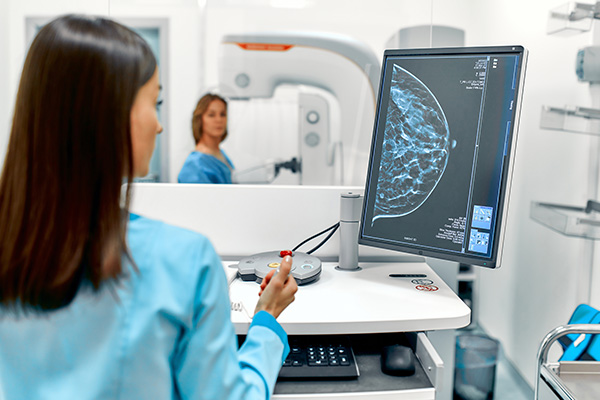
Digital Mammography
Digital mammography is a form of mammography that uses digital receptors and computers instead of x-ray film to help examine breast tissue for breast cancer. The electrical signals can be read on computer screens, permitting more use of images to allow radiologists a clear view of the results.
Breast Ultrasound
A breast ultrasound uses sound waves to create a picture of the tissues inside the breast. Unlike a mammogram, a breast ultrasound shows all areas of the breast, including the area closest to the chest wall, which is hard to study with a mammogram. A breast ultrasound should not replace a mammogram, but it is helpful to see whether a breast lump is filled with fluid, known as a cyst, or if it is a solid lump. It does not use x-rays or other potentially harmful types of radiation.

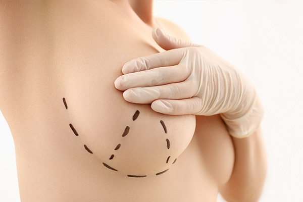
Breast Biopsy
A breast biopsy is a quick, accurate, and minimally invasive procedure that evaluates a suspicious area in the breast to determine if it is cancer. A small sample of breast tissue is removed for laboratory testing to identify and diagnose abnormalities in the cells to see if additional surgery or treatment is necessary.
Fine Needle Aspiration
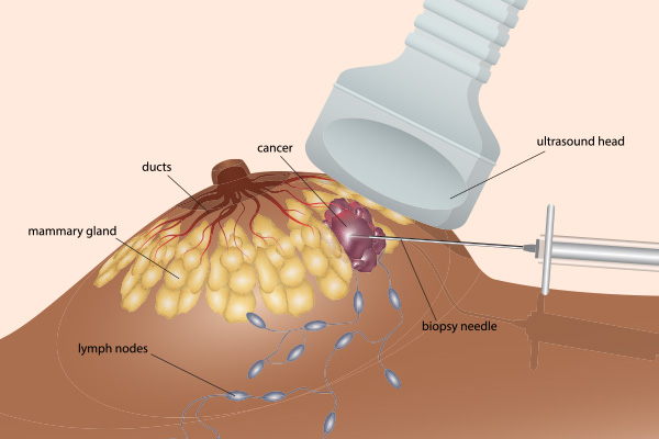
During a fine-needle aspiration, a thin needle is inserted into a breast lump to withdraw fluid. This procedure is often done using ultrasound to guide accurate placement of the needle. If the fluid comes out and your lump goes away successfully, your doctor can make a breast cyst diagnosis.
Cyst Aspiration
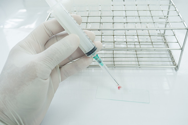
This is a simple procedure performed to drain a symptomatic or questionable cyst within the breast tissue. In this procedure, local anesthesia is used to numb the skin. Then the breast radiologist guides a thin needle into the cyst using ultrasound guidance. The fluid is then withdrawn into a syringe. The fluid is usually discarded (not tested) unless blood is found.
Breast MRI
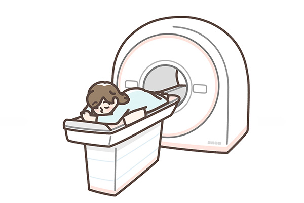
A Breast MRI is an imaging test that helps determine if a lump in the breast is cancerous or benign. This test uses a magnetic field and pulses of radio waves to make pictures of the breast. The Breast MRI may show problems in the breast that cannot be seen on a mammogram, ultrasound, or CT scan. The picture shows your breast’s normal structure, and identifies tissue damage or disease, inflammation, or a lump.

Genetic Testing for Hereditary Cancer Risk
Hereditary cancer is caused by an inherited genetic mutation. Approximately, 10-15% of most cancers are due to inherited genetic mutations. In these families, it is typical to see a recurring pattern of cancer across two to three generations—like multiple individuals diagnosed with the same type of cancer(s) and individuals diagnosed with cancer much younger than average.
A mutation can greatly increase your risk for developing cancer. Mutations in the genes that increase risk for cancer are not that common, but when present they significantly increase the chances someone might develop cancer in his or her lifetime.
For example, a BRCA1 mutation can increase a woman’s chance of breast cancer up to 81% and ovarian cancer up to 54% by age 80.
Your provider may adjust your screening plan if you have a mutation.
Knowing that you have a mutation that increases your risk of developing cancer allows you and your healthcare provider to create a personalized screening plan, which increases the chance of early detection.
The Diagnostic Center for Women is partnering with Color to offer comprehensive genetic testing for 8 different types of cancer (Breast, Ovarian, Colon, Uterine, Melanoma, Stomach, Pancreatic and Prostate) to any person who is interested in learning if they have an increased risk for hereditary cancer.

